 Pristupačnost
- Equipment Catalogue of the Faculty of Chemical Engineering and Technology
- XRD Shimadzu 6000
- SEM Tescan Vega 3
Equipment Catalogue of the Faculty of Chemical Engineering and Technology
Equipment Catalogue (pdf, 5.05 MB)
![]()
Apparatus: X-ray diffractometer
Manufacturer: Shimadzu
Short description of the method: Powder X-ray Diffraction (XRD) is a technique in which a beam of nearly monochromatic X-rays is directed onto the flat surface of a finely ground material placed in a sample holder. The sample-diffracted X-rays are measured in dependence on diffraction angle. The obtained data reveal information about the structure of the material(s) present in the sample.
Purpose: X-ray diffraction technique for powder samples is one of the most useful methods for exploring the structure of matter. The simplest application of powder XRD is phase identification (qualitative analysis). Quantitative XRD, the determination of the relative amounts of different phases in a given sample, is also possible, although in most cases it is a difficult undertaking, requiring time and considerable expertise. Other applications include the determination of crystal lattice parameters, the determination of crystallite size, degree of crystallization and strain analysis.
Technical characteristics: Basic components of the system are high-voltage generator, broad focus X-ray tube with CuKa radiation and Ni filter, high-precision vertical goniometer, graphite monochromator, scintillation counter and computer with system software.
Sample type and preparation: Solid sample, crystalline, inorganic/organic, naturally occurring/synthetic. The sample has to be milled to a fine powder.
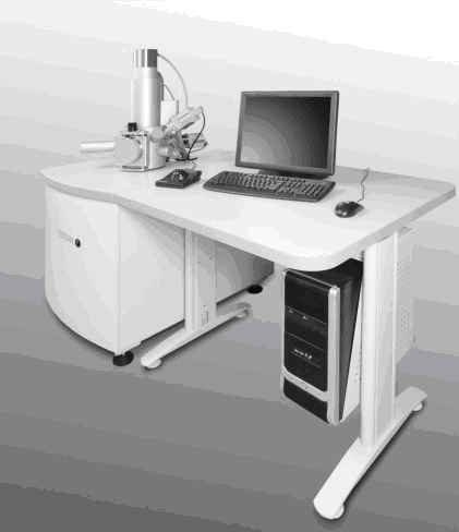
Apparatus: Scanning electron microscope
Manufacturer and type: Tescan Vega III Easyprobe
Short description of the method: Scanning electron microscopy (SEM) is an imaging technique in which specimen surface is scanned by a focused electron beam, resulting in an enlarged (up to 100 000 times) image of the specimen. Image contrast is dependant primarily on surface topography, and in case of backscattered electron (BSE) detection, on atomic number of atoms in the specimen. The microscope is also equipped with an energy dispersive X-ray spectroscopy (EDS) detector, which enables elemental analysis of the specimen.
Purpose: Scanning electron microscopy makes possible observation of dry specimens at high magnifications, and resolution of details that cannot be achieved in conventional optical microscopy, with relatively simple specimen preparation. It can be used to determine specimen morphology, particle and pore size, and also elemental composition if X-ray spectroscopy (EDS) is used.
Technical characteristics: Tescan Vega III Easyprobe, with tungsten filament, accelerating voltage range of 5 – 30 kV, magnification range of 100 – 100 000 times, maximum declared resolution of 3 nm, secondary (SE) and backscattered electron (BSE) detectors, as well as detector for energy dispersive X-ray spectroscopy (EDS).
Specimen type and preparation: Dry specimens of all types, powdery or with size ranging from under 1 cm to maximal 10 cm. Non-conductive specimens must first be sputter-coated with a conductive layer (sputter-coater is available). As the microscope requires high vacuum, specimen should not outgas under vacuum.